Algorithm that identifies heart-muscle thickening from routine heart activity test has been created with data from more than 37,500 UK Biobank participants.
Summary
An artificial-intelligence algorithm, created using data from more than 37,500 UK Biobank participants, can use a routine heart-activity test to predict dangerous heart-muscle thickening. This condition rarely causes symptoms but can lead to heart failure or cardiac arrest. Usually, a specialised heart scan is needed to identify it. Algorithms could help doctors to spot people who are at risk of heart disease early, while sparing people with a healthy heart unnecessary tests.
An artificial-intelligence (AI) algorithm can use a routine heart activity test to predict dangerous heart-muscle thickening, a condition that can currently only be identified with specialised scans. AI algorithms might eventually be able to help doctors to better identify those at risk of heart disease and put them forward for diagnostic scans – while sparing people with a healthy heart unnecessary tests.
Thickened muscles on the heart’s left side (known as left ventricular hypertrophy, or LVH) affects an estimated 15 to 20% of the population. LVH rarely causes symptoms, but can result in heart failure or even sudden cardiac arrest. Treating the underlying cause of LVH, high blood pressure for example, can keep that risk in check.
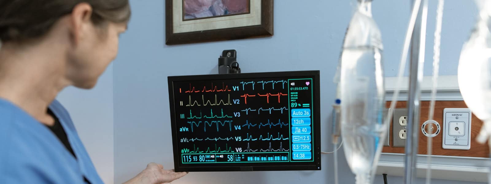
[An electrocardiogram (ECG)] is much easier, it’s quicker to do than an MRI, and it’s cheap and effective.
Professor Patricia Munroe, Queen Mary University of London, UK
People aren’t routinely screened for LVH because this usually means doing a heart ultrasound or an MRI (magnetic resonance imaging), for which there can be long waiting times. “[An electrocardiogram (ECG)] is much easier, it’s quicker to do than an MRI, and it’s cheap and effective,” explains the study’s co-leader Patricia Munroe from Queen Mary University of London, UK.
AI spots the difference
Munroe and her team trained and tested an AI algorithm on ECG data and measurements such as blood pressure of more than 37,500 UK Biobank participants. The participants’ heart imaging scans were used to check if they really had LVH or not.
The algorithm correctly identified 70% of people with LVH. This is a big step forward compared with the around 30% accuracy of other algorithms, says Munroe, and the team is working on improving this even further.
Identifying people with LVH “is already something that you can do, to some extent, just by looking at the ECG, but they were able to go much more in depth and be more accurate using the machine-learning technique”, comments Arunashis Sau from Imperial College London, UK, who also works on AI-enhanced ECG.
Some health conditions are apparent to clinicians in the size and shape of the wiggly ECG line – others can be too subtle for a human to spot. “A major part of what AI can do is connect all the different tiny little changes in an ECG,” Sau says.
The algorithm will need to be further refined and tested on a larger and more diverse group of people to get it ready for the clinic, Munroe suggests. It also needs to be cost-effective, Sau points out: “It needs to be increasing diagnoses without overburdening the healthcare system.”